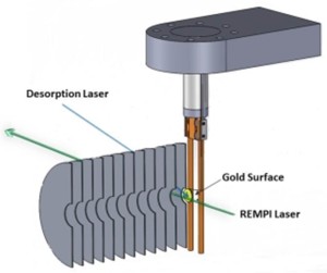
In adapting VMI for surface studies, we placed a single crystal of gold Au(111) in-plane with the first plate of the VMI optics (the repeller), adsorbed nitric oxide (NO), and laser-desorbed intact NO molecules using a pulsed UV laser.
The ejected neutral NO molecules are then ionised in a REMPI scheme 3mm above the surface, hence the time delay between the two lasers (desorption and ionisation) delivers time-of-flight (and hence) velocity information along the surface normal.
Together with the velocity distribution in the 2 dimensions parallel to the surface obtained from the velocity map images, we obtain fully resolved 3-dimensional velcity distributions of fragments ejected from surfaces, (one of) the holy grail in surface reaction dynamics.

|
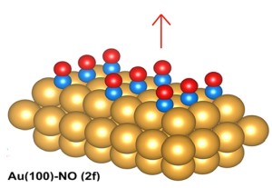
We could thus study the laser desorption of NO from gold and determine "the red arrow" in the right picture, i.e. the velocity distribtion of the NO in 3 dimensions independently. This is shown on the left where the speed distribution along the surface normal is vz, and the speed distributions in the two remaining dimensions parallel to the gold surface (vx and vy) are averaged to simplify graphical representation. |
|
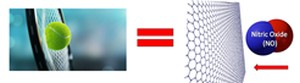
| We are currently studying the scattering of nitric oxide molecules, NO, off graphene surfaces supported by a gold substarte, in a way not too dissimilar to playing tennis with a racquet (the strings are the bonds in graphene) and a tennis ball (NO). |
|